

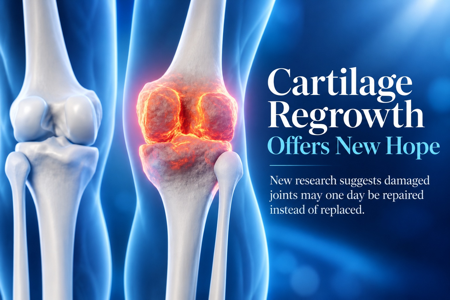
Scientists at Stanford Medicine have identified a promising approach that appears capable of regenerating worn joint cartilage by blocking an ageing-related protein known as 15-PGDH. In experiments involving older mice, the treatment restored cartilage that had naturally deteriorated with age and dramatically reduced the development of osteoarthritis following knee injuries similar to ACL tears. Even more encouragingly, human cartilage obtained from patients undergoing knee replacement surgery also began producing new articular cartilage when exposed to the treatment. While the research is still at an early stage, the findings raise the possibility that future medicines could one day repair damaged joints instead of simply managing pain until replacement surgery becomes necessary.
The research challenges the long-standing belief that adult articular cartilage has very little capacity to repair itself. Rather than relying on stem cells, the treatment appears to make existing cartilage cells behave more like younger cells again. If the same effect can eventually be reproduced safely in patients, researchers believe it could open the door to oral medicines or joint injections designed to regenerate cartilage directly.
Osteoarthritis Currently Has No Drug That Reverses The Damage
Osteoarthritis develops as the smooth cartilage covering the ends of bones gradually breaks down. As the protective tissue becomes thinner and softer, bones move less smoothly against one another, resulting in pain, swelling, stiffness and reduced mobility. The condition affects roughly one in five adults in the United States and is estimated to generate around US$65 billion in direct healthcare costs each year.
Existing treatments largely focus on controlling symptoms rather than repairing the underlying damage. Painkillers, anti-inflammatory medicines, physiotherapy and lifestyle changes may help patients remain active, but they do not reliably restore the cartilage that has already been lost. Once the joint becomes severely damaged, knee or hip replacement surgery may eventually become the only practical option.
That is what makes the Stanford research particularly interesting. Instead of simply reducing inflammation or pain, the scientists targeted a biological mechanism associated with ageing itself. The goal was to see whether older cartilage could be pushed back towards a healthier and more regenerative state.
The Key Target Is An Ageing-Related Protein Called 15-PGDH
The study focused on 15-PGDH, a protein that becomes increasingly abundant as tissues age. Researchers have described proteins such as this as "gerozymes", referring to enzymes that rise with age and contribute to the gradual decline in tissue function. Previous research from the same group had already shown that blocking 15-PGDH could improve regeneration in several other parts of the body.
Earlier experiments found that inhibiting the protein increased muscle mass and endurance in older mice. Increasing 15-PGDH levels in younger animals produced the opposite effect, causing muscles to shrink and weaken. The pathway has also been linked with regeneration involving bone, nerves, blood cells, the liver and other tissues.
The protein works partly by breaking down prostaglandin E2, or PGE2, a molecule involved in several biological processes. Previous studies showed that modestly increasing PGE2 by blocking 15-PGDH could support tissue repair. Researchers therefore wanted to know whether the same pathway might also influence the age-related deterioration of cartilage.
Older Mice Had Roughly Twice As Much 15-PGDH
When researchers compared knee cartilage from young and old mice, they found that levels of 15-PGDH were approximately twice as high in the older animals. That finding suggested the protein might be playing a role in the gradual decline of cartilage with age. The team then treated older mice with a small molecule designed to inhibit 15-PGDH.
The drug was tested in two different ways. In one experiment, it was injected into the abdomen so the treatment could circulate throughout the body. In another, researchers delivered it directly into the affected knee joint.
Both approaches produced surprisingly strong results. Cartilage that had become thin and degraded with age began growing thicker across the joint surface after treatment. More importantly, the regenerated tissue was the type of cartilage joints actually need to function properly.
The Treatment Regenerated Hyaline Cartilage
There are several different types of cartilage in the human body, and they are not interchangeable. Elastic cartilage provides flexible support in structures such as the outer ear, while fibrocartilage is tougher and designed to absorb force in areas including the spine. Hyaline cartilage is smoother and allows bones to glide against each other with minimal friction.
Within joints, this smooth tissue is known as articular cartilage, and it is the type most commonly destroyed by osteoarthritis. Researchers were therefore particularly interested in whether the treatment would restore proper hyaline cartilage rather than producing fibrocartilage, which would not provide the same smooth joint surface. The treated mice produced the desired hyaline cartilage.
Nidhi Bhutani, an associate professor of orthopaedic surgery and one of the study's senior authors, described the degree of regeneration in aged animals as remarkable. The result was particularly unexpected because adult articular cartilage has traditionally been considered very poor at repairing itself once damaged.
Existing Cartilage Cells Appeared To Become Younger
Researchers initially expected that stem or progenitor cells might be responsible for the regeneration. In many tissues, repair occurs when specialised stem cells multiply and then develop into the cells needed to replace damaged structures. Surprisingly, that was not what appeared to be happening inside the cartilage.
Instead, existing cartilage cells known as chondrocytes changed their patterns of gene activity. Older chondrocytes normally showed increased activity in genes associated with inflammation, tissue breakdown and the unwanted transformation of cartilage towards bone. Genes involved in producing and maintaining healthy cartilage were comparatively less active.
After 15-PGDH inhibition, that balance changed substantially. The existing cells shifted towards a gene-expression pattern associated with healthier and younger cartilage rather than being replaced by a new population of stem cells. According to senior author Helen Blau, this represents a different approach to adult tissue regeneration and could potentially have significant clinical importance.
The Cellular Changes Were Dramatic
One population of older chondrocytes that produced 15-PGDH and expressed genes associated with cartilage degradation fell from around 8% of cells to 3% after treatment. Another population associated with fibrocartilage formation decreased from approximately 16% to 8%. These reductions indicated that fewer cells were behaving in ways linked to ageing and deterioration.
At the same time, the proportion of cells associated with healthy hyaline cartilage moved sharply in the opposite direction. Chondrocytes that did not produce 15-PGDH and expressed genes involved in creating hyaline cartilage and maintaining the extracellular matrix increased from 22% to 42%. The extracellular matrix is particularly important in cartilage because it provides the structural environment that allows the tissue to withstand pressure while maintaining a smooth joint surface.
Taken together, the findings suggest that inhibiting 15-PGDH did not simply slow cartilage loss. It appeared to reprogramme the behaviour of existing cells so that the tissue began acting biologically younger and rebuilding itself.
Treatment Also Protected Mice After ACL-Like Injuries
The researchers also investigated whether the approach could prevent osteoarthritis following traumatic knee injuries. ACL injuries are particularly relevant because they are common in sports involving rapid direction changes, jumping or sudden stops. Even when the damaged ligament is surgically repaired, the injury can have long-term consequences for the joint.
Around half of people who experience these types of injuries eventually develop osteoarthritis in the affected knee within approximately 15 years. This means repairing the ligament does not necessarily prevent progressive cartilage damage later in life. Researchers wanted to know whether blocking 15-PGDH could interrupt that process.
Mice received the inhibitor twice a week for four weeks after their knees were injured. The treatment dramatically reduced their likelihood of developing osteoarthritis, while control animals developed the disease within four weeks. The untreated injured animals also showed approximately twice the 15-PGDH levels seen in uninjured knees.
Treated Animals Also Moved More Normally
The benefit was not limited to what researchers observed under a microscope. Animals receiving the treatment moved more normally and placed more weight on the paw connected to the injured leg. This suggests that the cartilage improvements translated into functional differences in how the animals used the affected joint.
The findings are especially interesting because prostaglandin E2 is commonly associated with inflammation and pain. However, the researchers found that relatively small increases at normal biological levels could instead help stimulate regeneration. This demonstrates how the same signalling molecule can have very different effects depending on its concentration and biological context.
If similar protection could eventually be achieved in people, treatment after a serious knee injury might one day involve not only repairing damaged ligaments but also protecting the cartilage from the progressive deterioration that can follow years later.
Human Cartilage Responded To The Treatment Too
Perhaps the most encouraging part of the study involved cartilage collected from people with osteoarthritis who were undergoing total knee replacement surgery. Researchers exposed these human tissue samples to the 15-PGDH inhibitor for one week. The results showed changes broadly consistent with what had been observed in mice.
The treated human samples contained fewer chondrocytes producing 15-PGDH. Genes associated with cartilage degradation and fibrocartilage formation also became less active compared with untreated samples. Most notably, the tissue began regenerating articular cartilage.
This does not yet prove that the same treatment will regrow cartilage inside a living human knee. Laboratory-treated tissue and animal experiments are important steps, but human joints are considerably more complex and clinical trials will still be required. Nevertheless, seeing human osteoarthritis tissue respond in a similar direction strengthens the case for moving the research towards patient studies.
A 15-PGDH Drug Has Already Reached Human Testing For Another Condition
There is another encouraging piece of the story: oral 15-PGDH inhibition is not entirely untested in humans. A drug targeting the same pathway has already entered Phase 1 clinical trials for age-related muscle weakness. According to Blau, those trials showed that the inhibitor was safe and biologically active in healthy volunteers.
That does not mean the drug has already been proven safe or effective for cartilage regeneration. Dosage, treatment duration, delivery method and the biology of damaged joints may require different approaches. A dedicated clinical trial would still need to determine whether cartilage can be regenerated safely and meaningfully in patients with osteoarthritis.
The researchers hope such a trial can begin in the future. Their longer-term vision is ambitious: rather than waiting until a damaged knee or hip requires replacement, doctors might eventually be able to stimulate the patient's own remaining cartilage to rebuild itself.
Could This Eventually Reduce The Need For Joint Replacement?
It is far too early to say that knee or hip replacement surgery is on the way out. Joint replacement remains an extremely effective treatment for severe osteoarthritis, and the new approach has not yet demonstrated that it can restore an arthritic human joint inside the body. The current evidence comes primarily from mice and human cartilage treated outside the body.
However, the research changes an important assumption about cartilage biology. Articular cartilage may not be as permanently incapable of regeneration as previously believed. If older chondrocytes can be shifted back towards a healthier state, the therapeutic possibilities extend beyond simply slowing deterioration.
Future treatments could potentially involve an oral medicine, a targeted injection into the joint or some combination of approaches. Instead of replacing an entire joint after cartilage has disappeared, treatment might eventually focus on preserving and regenerating the tissue before damage becomes irreversible.
Important Research Disclosures
The work involved researchers from Stanford Medicine and the Sanford Burnham Prebys Medical Discovery Institute and received funding from several research organisations and foundations. The research was published in Science and involved specialists in stem-cell biology, orthopaedic surgery and ageing biology.
Several researchers involved in the study are also named as inventors on patent applications covering the use of 15-PGDH inhibition for cartilage and tissue rejuvenation. Those patents are held by Stanford University and licensed to Epirium Bio. Blau is also a cofounder of Myoforte/Epirium and holds financial interests in the company.
These disclosures do not invalidate the scientific findings, but they are relevant when considering how the research may eventually progress towards commercial therapies. Independent clinical testing will ultimately be necessary to establish whether the treatment delivers meaningful benefits to patients.
Final Thoughts
The Stanford-led research offers one of the more intriguing approaches yet to the problem of cartilage regeneration. By blocking the ageing-related protein 15-PGDH, scientists were able to restore substantial amounts of healthy articular cartilage in older mice, reduce osteoarthritis after knee injury and push ageing cartilage cells towards a younger biological state. The fact that human osteoarthritis tissue also began producing new cartilage after treatment makes the findings particularly encouraging.
The important caveat is that this is not yet a cure for osteoarthritis and certainly not the end of knee or hip replacement surgery. Dedicated human clinical trials still need to determine whether the effect can be safely reproduced inside patients and whether regenerated cartilage provides lasting improvements in pain and mobility. Even so, the possibility of encouraging existing cartilage to rebuild itself represents a major shift in how degenerative joint disease might eventually be treated.











Comments 0